
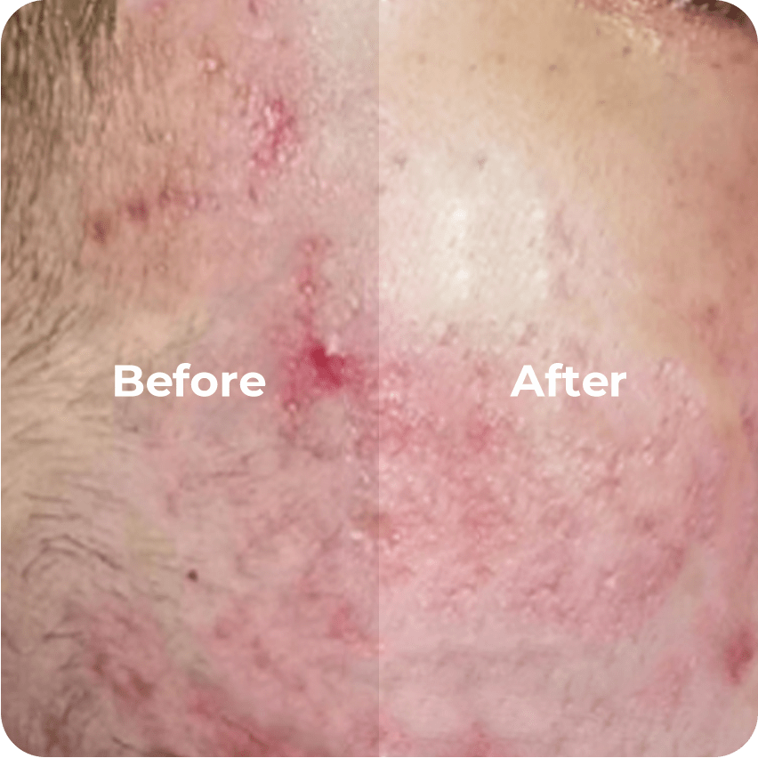
Why a comparative view helps clinics and patients
Please allow a short comparison to clarify choices when planning a pigmentation removal treatment. Different laser systems trade off wavelength, pulse width, and energy delivery, and those trade-offs directly affect outcomes, downtime, and risk of hypo- or hyperpigmentation. In many clinics—both in Tokyo dermatology centers and large U.S. academic hospitals—teams select devices after balancing efficacy against skin-type safety. Regulatory bodies such as the U.S. Food and Drug Administration (FDA) have cleared several lasers for pigmented lesion treatment, which gives an important real-world anchor for clinical practice and device selection.
Core parameters to compare
When comparing systems, three parameters most strongly influence treatment behaviour: wavelength, pulse width, and fluence. Wavelength determines chromophore targeting (melanin vs. other absorbers). Pulse width (or pulse duration) sets how quickly energy is delivered; shorter pulses tend to confine energy to pigment granules, while longer pulses allow heat diffusion into surrounding tissue. Fluence (energy per area) and spot size complete the practical set — they together shape clinical endpoint and risk profile. Understanding these terms supports safer protocol design and more predictable results.
Modalities at a glance: strengths and limits
It is useful to compare common options side by side. Below are concise notes to guide selection, with emphasis on practical differences.
- Q‑switched Nd:YAG (532/1064 nm): Effective for many epidermal and dermal pigmented lesions. Good for darker inks and deeper pigment with 1064 nm; 532 nm targets superficial melanin. Advantage: established safety record. Limitation: multiple sessions often required; risk of post‑inflammatory pigment change on darker Fitzpatrick skin types.
- Picosecond lasers: Shorter pulse widths provide stronger photomechanical effect on pigment, often needing fewer sessions. Advantage: improved clearance for certain lentigines and tattoos. Limitation: cost and variable evidence for all pigment types.
- Fractional (ablative/non‑ablative) resurfacing: Combines microthermal injury with pigment removal and stimulates remodeling. Advantage: treats blended concerns (texture + pigment). Limitation: greater downtime and wound‑care requirements.
- Intense Pulsed Light (IPL): Broad-spectrum, adjustable filters for superficial pigmented lesions. Advantage: cost-effective for epidermal pigment. Limitation: less precise than lasers; operator-dependent outcomes.
For clinics seeking a precise option labeled as laser treatment to remove pigmentation, the decision often rests on patient skin type, lesion depth, and tolerance for multiple sessions.
Practical protocol considerations and common mistakes
In practice, please consider the following procedural priorities. First, always document Fitzpatrick skin type and perform a test spot on the least visible area. Second, cooling strategy and pulse overlap must be specified in the protocol — inadequate cooling or excessive overlap is a common source of burns. Third, realistic endpoint cues should be defined (e.g., immediate lightening, mild whitening, or subtle erythema), so the operator knows when to stop increasing fluence. A frequent mistake is treating to maximal fluence without considering cumulative thermal load — this can cause paradoxical darkening, especially in higher‑melanin skin. — It is prudent to start conservatively and escalate across sessions rather than risk a single aggressive treatment.
Device selection checklist for clinics
Please review these operational items before purchase or protocol adoption:
- Evidence base: peer-reviewed outcomes for the specific pigment types you treat.
- Adjustability: ability to vary wavelength, pulse width, spot size, and fluence with fine increments.
- Safety features: integrated cooling, real-time energy monitoring, and documented maintenance/service plans.
- Training and support: manufacturer or distributor training for staff and access to clinical protocols for different Fitzpatrick types.
Advisory: three golden rules for selecting and using systems
1) Prioritize match of wavelength to lesion depth: choose 532 nm or IPL filters for superficial epidermal pigment, and 1064 nm for deeper dermal pigment. This improves clearance and reduces collateral damage. 2) Control pulse width relative to target granule interaction: when available, shorter pulses (picosecond) can reduce sessions for fragmented pigment, but require precise parameter control. 3) Measure safety by outcomes, not promises: evaluate a device by clinic‑level metrics — clearance rate at 3–6 months, incidence of post‑inflammatory hypo/hyperpigmentation, and number of sessions per case. These metrics will indicate whether a system performs as claimed.
Final reflection and how ENZOEYS fits
Comparative selection is both science and workflow design. Clinics that align wavelength, pulse width, and cooling protocols to patient factors tend to achieve repeatable results with fewer adverse events. ENZOEYS integrates evidence‑based parameter sets, operator training, and practical device configurations to help teams deliver measured improvements in pigmentation care — a natural fit for clinics prioritizing consistent, patient‑safe outcomes. —